Banque d`images

In the peripheral blood (May-Grünwald-Giemsa stain) of this patient lymphoma cells can be detected. They show a large, deeply indented nucleus (->) and scanty cytoplasm. Further diagnostics confirmed a mantle cell lymphoma.
<p>In the peripheral blood (May-Grünwald-Giemsa stain) of this patient lymphoma cells can be detected. They show a large, deeply indented nucleus (->) and scanty cytoplasm. Further diagnostics confirmed a mantle cell lymphoma.</p>

Peripheral blood (May-Grünwald-Giemsa stain) of an 82-year old patient with RCMD showing a macrocytic, hyporegenerative anaemia with clear changes of the red blood cells (micro- and macrocytosis, poikilocytosis and some teardrop cells (->)).
<p>Peripheral blood (May-Grünwald-Giemsa stain) of an 82-year old patient with RCMD showing a macrocytic, hyporegenerative anaemia with clear changes of the red blood cells (micro- and macrocytosis, poikilocytosis and some teardrop cells (->)).</p>

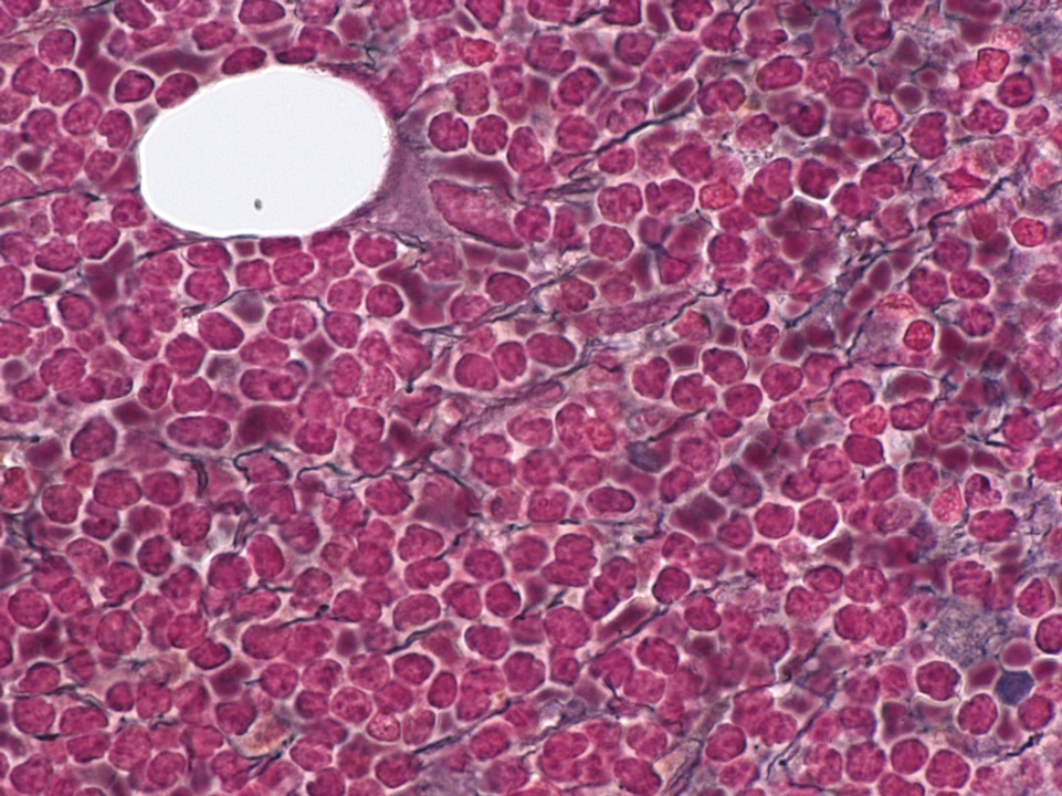
Bone marrow histology (Gomori stain) of a patient showing an infiltration of mantle cell lymphoma cells and an increase in fibres (black lines).
<p>Bone marrow histology (Gomori stain) of a patient showing an infiltration of mantle cell lymphoma cells and an increase in fibres (black lines).</p>

May-Hegglin anomaly belongs to a family of macrothrombocytopenias characterised by mutations in the MYH9 gene. It is a rare autosomal dominant disorder characterised by various degrees of thrombocytopenia that may be associated with purpura and bleeding.
<p>May-Hegglin anomaly belongs to a family of macrothrombocytopenias characterised by mutations in the MYH9 gene. It is a rare autosomal dominant disorder characterised by various degrees of thrombocytopenia that may be associated with purpura and bleeding. </p>

Cell description:
Size: 10-12 µm
Nucleus: kidney or U-shaped with clumped chromatin
Cytoplasm: acidophilic
neutrophil: fine reddish granulation
Cell division is not possible anymore and protein synthesis has stopped.
<p>Cell description: </p> <p>Size: 10-12 µm </p> <p>Nucleus: kidney or U-shaped with clumped chromatin </p> <p>Cytoplasm: acidophilic </p> <p>neutrophil: fine reddish granulation </p> <p>Cell division is not possible anymore and protein synthesis has stopped.</p>

Blood film prepared after storage of the EDTA blood for more than one day. A safe morphological differentiation is no longer possible.
<p>Blood film prepared after storage of the EDTA blood for more than one day. A safe morphological differentiation is no longer possible.</p>

Cell description:
Size: 20 µm
Nucleus: kidney- to band-shaped
Cytoplasm: grey and clear with fine azurophilic granules
They are shortly located in the peripheral blood and then move into the tissue where they differentiate into macrophages. Function: Phagocytosis either of harmful pathogens or dead, dying or damaged cells from the blood.
<p>Cell description: </p> <p>Size: 20 µm </p> <p>Nucleus: kidney- to band-shaped </p> <p>Cytoplasm: grey and clear with fine azurophilic granules </p> <p>They are shortly located in the peripheral blood and then move into the tissue where they differentiate into macrophages. Function: Phagocytosis either of harmful pathogens or dead, dying or damaged cells from the blood. </p>

